Microscopy and analysis

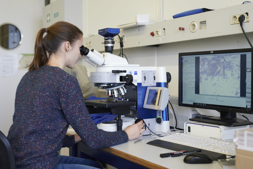
The institute WWI is equipped with many modern microscopic methods for the microstructural characterization of samples. In addition to light optical microscopy, scanning electron microscopy (SEM) and transmission electron microscopy (TEM) are of particular importance. The focused ion beam (FIB) technique on the SEM also allows targeted analysis of interesting sample sites. A corresponding analysis allows the determination of the chemical composition (EDX) and the crystallographic orientation (EBSD). A laser scanning microscope (shared device) and an atomic force microscope are also available for the high-resolution examination of surface structures. By means of the available X-ray diffractometers, the phases occurring in the materials can be analyzed (e.g. crystal structure, lattice parameters, texture, …). A heating chamber allows investigations up to 1100°C. Particularly noteworthy are the two atom probes, which allow the spatially resolved chemical composition of an interesting sample range to be determined with atomic resolution.
- Atom probe – Cameca Leap 4000X HR
- Atom probe – Oxcart
- Dynamic differential calorimetry – Netsch 204 F1 Phoenix®
- Field ion microscope
- Large chamber scanning electron microscope (LC-SEM)
- Scanning electron microscope with FIB- FEI Helios NanoLab 600i DualBeam
- Scanning electron microscope with FIB – Zeiss Crossbeam 1540
- Atomic force microscope – Dimension 3100
- X-Ray diffractometer- Bruker D5000
- Röntgendiffraktometer – Bruker D8
- Transmission electron microscope – Philips CM200