Nanoparticles in the atom probe

Outstanding potential of nanoparticle (NP) atom probe tomography (APT) analysis has been obvious from the first publication [1]. Still, data fidelity has not reached up to its potential of 3D structural and chemical analysis with atomic resolution [2]. One of the main reasons for this is that sample fabrication is challenging: Starting with a sample being smaller than an APT sample tip (100 nm diameter) requires additive production approaches rather than the well-established substractive production approaches (e.g. FIB-lift-out [3]). A recent publication stated[4], there are just limited reports about successful NP APT experiments. Most of them introduced their own sample fabrication method.
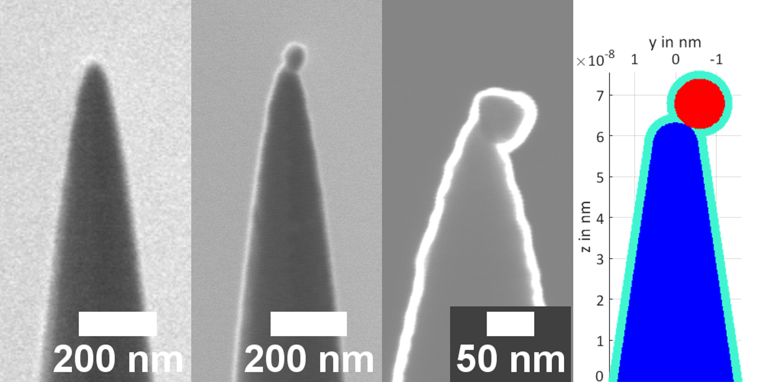
In this project we are working on methods with various approaches for sample NP APT sample fabrication. The most promising combines an in-situ SEM lift-out strategy [5] with a specialised electron beam – physical vapor deposition deposition (EB-PVD). Different stages during this method are presented in Fig. 1. Custom build instruments, mostly ultra-high-vacuum instruments, were built and are successivley improved. Experimental data is analysed supported by 3D-full-scale APT simulation [6].
[1] Tedsree, K., et al. (2011) Nat. Nano-technol.
[2] Felfer, P., et al. (2015) Ultramicroscopy
[3] Thompson, T. (2007) Ultramicroscopy
[4] Kim, S.-H., et al. (2020) J. Alloys Compd.
[5] Przybilla, T., et al. (2018) Small Methods
[6] Oberdorfer, C., et al. (2013) Ultramicroscopy